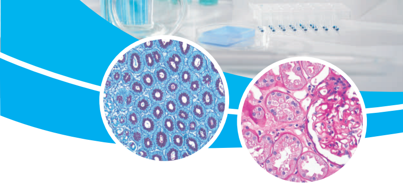
Special Stains For Automatic and Manual use

INTRODUCTION
Special Staining is the most routinely used term to designate stains that allow the recognition of specific chemical groups in the histological structure of tissues and, consequently, to identify their nature on numerous occasions. These histochemical stains are based on the different affinity of the cellular components to fix the chemical groups present in the different staining solutions and constitute complementary tools of the first order for routine staining, so that for many years they have become essential in daily anatomopathological practice. In this sense, the presence of deviations from normal staining in terms of location, quantity, intensity, structure, etc.
Is associated with numerous specific pathological processes and, likewise, the particular and anomalous location of some chemical groups can identify specific cell types and various pathogenic microorganisms. Due to the fact that the correct assessment of histochemical techniques requires strictly following their technical protocol, which is often quite complicated, with the consequent lack of standardization and reproducibility that always leads to a high degree of uncertainty in the interpretation of histochemical techniques by the pathologist.
For this reason, its adaptation, optimization and validation both manually and in an automatic staining instrument as versatile and safe as the MD Stainer contributes to granting a high degree of reproducibility to the processes and likewise provides the Laboratory Technician with great comfort and soundness in handling staining protocols. All the procedures included here have been designed, adapted and validated to be used manually and/or in MD Stainer
